The article summarized below describes a new way to help the more than 35 million TMJ patients with TMJ disc degeneration and mechanical dysfunction, symptoms which can lead to surgery in 10 percent of cases. TMJ discectomy/removal has been used in clinical practice for decades, but often with unsatisfactory outcomes. The potential of using a graft of a TMJ disc from a healthy donor has been largely unexplored due to the disc’s complicated structure and function, its limited availability, and inherent challenges in preserving the tissue including long-term storage options (greater than 6 months). The lack of research on donated TMJ discs has forced the use of replacement grafts from the patient’s own tissues, which can result in pain and functional loss, additional surgical sites, prolonged recovery times and elevated risk of surgical site infections. This study addresses an urgent need for providing “off-the-shelf” availability of grafts, for both TMJ discs and knee menisci, that possess natural properties closely resembling those of the native tissues.
The following article appeared in Clemson News on April 15, 2024 https://news.clemson.edu/cool-research-breakthrough-tissue-preservation-method-offers-new-hope-for-knee-and-jaw-patients/
New hope for millions of patients who suffer from knee and jaw problems is emerging from a study that came out of Clemson University’s partnership with the Medical University of South Carolina and an industry collaborator.
Researchers have found a promising new technique that could help stem the shortage of meniscus grafts. The grafts are often needed to treat injuries and degeneration in the knee or the jaw’s temporomandibular joint (TMJ).
The solution involves preserving donated meniscus tissue by freezing it in a process called vitrification. This process allows better storage of grafts for longer periods of time, making more tissue available for transplantation.
The research team was headed by Shangping Wang, an assistant professor of bioengineering at Clemson University. The team reported its findings in the journal Advanced Healthcare Materials.
The challenge: The meniscus is a rubbery, crescent-shaped tissue that cushions the joint and keeps it stable during movement, such as running or chewing. When the meniscus wears out or is injured, it has limited ability to repair itself and causes severe pain and difficulty moving.
Doctors often replace a damaged meniscus in a person’s knee or jaw joint with preserved meniscus tissue. But the supply of fresh meniscus tissue falls short of what is needed for the more than 1 million patients who undergo surgical repair or meniscectomy each year.
The primary obstacle is that viable meniscus tissues can be preserved for less than a month, leading to a critical shortage.
In the new study, researchers have proposed a promising solution known as vitrification. The process enables living cells to be chemically treated and then cooled to extremely low temperatures without harming the living cells.
Using vitrification, researchers are able to surround the tissue in a transparent, glassy state. The process effectively prevents the formation of damaging ice crystals within the cells of the tissue.
One of the challenges the scientists faced, though, is that meniscus tissues have dense structures that can be difficult to permeate. It can be challenging to penetrate the tissue with chemicals for preservation, and the tissue becomes exposed to the chemicals for too long.
When that happens, it can harm or kill the cells. To make vitrification work, it is critical to strike a balance between penetrating the tissue with chemicals but not exposing it for too long, researchers said.
The solution: To address the problem, researchers combined computational modeling with micro-CT imaging to pinpoint the crucial balance they wanted to achieve. They were able to determine the minimal exposure time necessary to effectively penetrate the tissue during vitrification, while avoiding damaging or killing the tissue.
The researchers then used what they found in the computational model to effectively preserve whole meniscus tissues both in knee and jaw joints.
The upshot of the study is that the vitrified meniscus tissues can be stored at very low temperatures for a long time, making the tissue suitable for transplantation.
“This pioneering work represents the first successful application of vitrification strategies to preserve whole viable grafts in both tissue types which opens new avenues for the successful preservation of various avascular tissues and holds the promise of facilitating widespread adoption of viable graft transplantation and research endeavors,” researchers wrote.
The title of the study is “Viable Vitreous Grafts of Whole Porcine Menisci for Transplant in the Knee and Temporomandibular Joints.”
A provisional patent stemming from the work has been established through the Clemson University Research Foundation, with backing from Executive Director Chris Gesswein.
The Musculoskeletal Transplant Foundation was a partial funding source for Wang’s research.
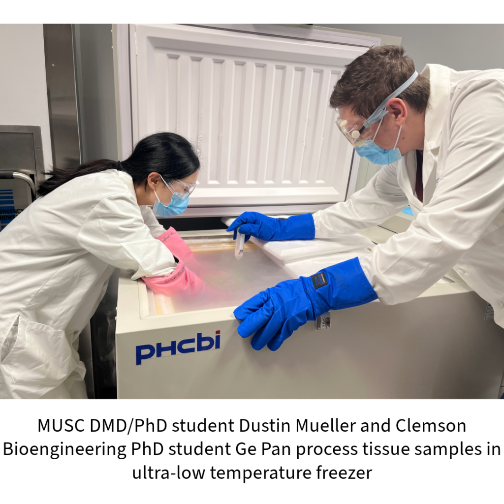
Equally contributing authors of the study are Dustin Mueller, a DMD/PhD student from the NIH T32 dental research training program within the Clemson-MUSC Bioengineering Program and Hai Yao, Clemson’s associate vice president for biomedical Innovation.
it is great news. my 42 years daughter is sufferting from tdj disorder and the disc is worn out. hopefully you will find a solution
Very helpful and informative. I appreciate all that you all do for people and what they have had to insure.
I would love to hear more about this and where I would go to receive this. 7 weeks ago I went in for arthroplasty on my TMJ articular disc. When the OMF surgeon got in, he found the disc had a hole in it. He put synvisc in the place of the disc and told me to pray. My follow-up is this week. I know I have to do orthodontics. Again, I would love to hear more about this.